Spinal Cord Injury Model Systems Consumer Information
Skin Care & Pressure Sores
Skin Care & Pressure Sores
• Part 1: Causes & Risks of Pressure Sores
• Part 2: Preventing Pressure Sores
• Part 3: Recognizing & Treating Pressure Sores
[Click here for a printer-friendly version of this page.]
Stages of pressure sores
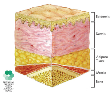
NORMAL SKIN
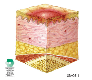
STAGE 1
Skin is not broken but is red or discolored. When you press on it, it stays red and does not lighten or turn white (blanch). The redness or change in color does not fade within 30 minutes after pressure is removed.
Click here to see photo: Stage 1
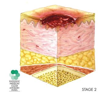
STAGE 2
The topmost layer of skin (epidermis) is broken, creating a shallow open sore. The second layer of skin (dermis) may also be broken. Drainage may or may not be present.
Click here to see photo: Stage 2
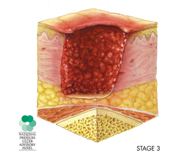
STAGE 3
The wound extends through the dermis (second layer of skin) into the subcutaneous (below the skin) fat tissue. Bone, tendon and muscle are not visible. Look for signs of infection (pus, drainage) and possible necrosis (black, dead tissue).
Click here to see photo: Stage 3
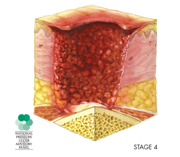
STAGE 4
The wound extends into the muscle and can extend as far down as the bone. Usually lots of dead tissue and drainage are present. There is a high possibility of infection.
Click here to see photo: Stage 4
SUSPECTED DEEP TISSUE INJURY
- Purple or maroon localized area of discolored intact skin or blood-filled blister due to damage of underlying soft tissue from pressure and/or shear. The area may be surrounded by tissue that is painful, firm, mushy, boggy, warmer or cooler as compared to nearby tissue.
- Deep tissue injury may be difficult to detect in individuals with dark skin tones. Progression may include a thin blister over a dark wound bed. The wound may further evolve and become covered by thin eschar (scab). Progression may be rapid exposing additional layers of tissue even with optimal treatment.
Click here to see photo: Deep tissue injury
Reference
Pressure Ulcer Stages Revised, by National Pressure Ulcer Advisory Panel (2007). <http://www.npuap.org>.
[Return to Part 3: Recognizing and Treating Pressure Sores]
Copyright and source information
©2009 Model Systems Knowledge Translation Center (MSKTC). This publication was produced by the SCI Model Systems in collaboration with the Model Systems Knowledge Translation Center (http://msktc. washington.edu) with funding from the National Institute on Disability and Rehabilitation Research in the U.S. Department of Education, grant no. H133A060070.
Our health information content is based on research evidence and/or professional consensus and has been reviewed and approved by an editorial team of experts from the SCI Model Systems.

