


SCI Pamphlets: Staying Healthy after a Spinal Cord Injury
The pamphlet series:
• Taking Care of Pressure Sores
• Maintaining Healthy Skin (Part I)
• Maintaining Healthy Skin (Part II)
• Taking Care of Your Bowels: The Basics
• Taking Care of Your Bowels: Ensuring Success
Bladder Management
[Download this pamphlet: "Bladder Management" - (PDF, 499KB)]
The Urinary System
The Urinary System is made up of five major parts:
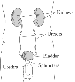
The Kidneys
The two kidneys filter waste and excess water from the blood and produce urine. Urine is being produced every minute of the day.
The Ureters
Each kidney has a thin, hollow tube that connects to the bladder. Urine flows down the ureters from the kidneys and empties into the bladder. The ureters have one-way valves in them, so even if you were to stand on your head urine could not flow back to the kidneys from the bladder.
The Bladder
The bladder is a collapsible sac lying in the pelvis. It is able to stretch to hold urine until you are ready to urinate. The bladder walls are made up of a series of muscles known collectively as the detrusor muscles. When you are ready to urinate, the detrusor muscles contract (squeeze) to help push the urine from the bladder. The lower portion of the bladder, which funnels urine into the urethra, is called the bladder neck or bladder outlet.
The Sphincter Muscles
The internal and external sphincter muscles form a ring around the urethra to keep urine in the bladder. When you are ready to urinate, these muscles relax to allow urine to flow out of the bladder.
The Urethra
The urethra is a small tube that allows urine to flow from the bladder to outside the body. The male urethra is 8-10 inches long and the female urethra is 1-2 inches long. The external urethral opening from the body is called the meatus for both men and women.
Voiding (Urination)
Normally, when the bladder becomes full (about 1-2 cups for most people), nerve endings in the bladder wall send a message to the brain via the spinal cord. The brain sends a message back to the bladder to contract the detrusor muscles and relax the sphincter muscles so you can void. If you can't get to a toilet, the brain delays the messages until you are ready to void.
After Spinal Cord Injury
The bladder, along with the rest of the body, undergoes dramatic changes. Since messages between the bladder and the brain cannot travel up and down the spinal cord, the voiding pattern described above is not possible.
Depending on your type of spinal cord injury, your bladder may become either "floppy" (flaccid) or "hyperactive" (spastic or reflex).
The Flaccid Bladder
A floppy bladder loses detrusor muscle tone (strength) and does not contract for emptying. This type of bladder can be easily overstretched with too much urine, which can damage the bladder wall and increase the risk of infection.
Emptying the flaccid bladder can be done with techniques such as Credé, Valsalva, or intermittent catheterization. It is very important that you do not let your bladder get overfull, even if it means waking up at night to catheterize yourself more frequently.
The Reflex Bladder
The detrusor muscles in a hyperactive bladder may have increased tone, and may contract automatically, causing incontinence (accidental voiding). Sometimes the bladder sphincters do not coordinate properly with the detrusor muscles, and medication or surgery may be helpful.
Bladder Management
Foley or Suprapubic Catheter
A tube is inserted through the urethra or abdomen and into the bladder, where a balloon on the end holds it in place. It remains in the bladder and drains constantly, so the bladder is never full.
Intermittent Catheterization
You drain your bladder several times a day by inserting a small rubber or plastic tube. The tube does not stay in the bladder between catheterizations.
Spontaneous Voiding
The bladder muscles contract to start the bladder-emptying process. This may be under your control (voluntary) or not (involuntary):
- Normal Voiding
This is done under your control. When the bladder gets full, messages are sent to the sacral level of the spinal cord and carried to the brain. The brain sends messages back to the bladder to contract, and to the sphincter muscle to open, so you can void. - Spincterotomy
This surgical process weakens the bladder neck and sphincter muscle to allow urine to flow out more easily. After this surgery, you will urinate involuntarily, and must wear a collection device. - Condom Catheter
These collection devices are worn by men for incontinence problems or after sphincterotomy (see above). They are made of latex rubber or silicone that covers the penis and attaches to a tube that drains into a collection bag.
Stimulated Voiding
Voiding is encouraged in one of several ways, such as:
- Anal or Rectal Stretch
This method for relaxing the urinary sphincter is usually used along with an abdominal corset and valsalva (see below). - Credé
This method involves manually pressing down on the bladder. - Tapping
The area over the bladder is tapped with the fingertips or the side of the hand, lightly and repeatedly, to stimulate detrusor muscle contractions and voiding. - Valsalva
This method involves increasing pressure inside the abdomen by bearing down as if you were going to have a bowel movement.
Surgical Alternatives
- Mitrofanoff
A passageway is constructed using the appendix so that catheterization can be done through the abdomen to the bladder. - Bladder Augmentation
Surgical enlargement of the bladder. - Spincterotomy
See the description of this procedure in the "Spontaneous Voiding" section.
University of Washington-operated SCI Clinics:
Harborview Medical Center
Rehabilitation Medicine Clinic
325 9th Ave., Seattle WA 98104
Spinal Cord Injury Clinic nurses: 206-744-5862
University of Washington Medical Center
Rehabilitation Medicine Clinic
1959 NE Pacific, Seattle WA 98195
Spinal Cord Injury Clinic nurses: 206-598-4295